Neuroscientific work
As exemplary
for the neuroscientific work we will present an eye movement recording system.
We will also offer insight into the research of the Max Planck Institute for
Brain Research, Fankfurt/Main. The works are thematically divided into seeing
and not-seeing. Further scientific contributions are outlined under www.locked-in.com.
 |
The eye movement recording system makes a representation of the gaze possible. Such an insight is, under normal circumstances, inaccessible. Our gaze is always moving and this sytem records its motions. The eye movement recording system tails our gaze while, for example we view ourselves. (Wolfgang H. Zangemeister and Ulrich Oechsner, "Infrared Recordings of Eye position and Eye fixation", Dept. of Neurology, University of Hamburg, eye movement recording system XY 1000, Eyetrace Inc., Sweden) |
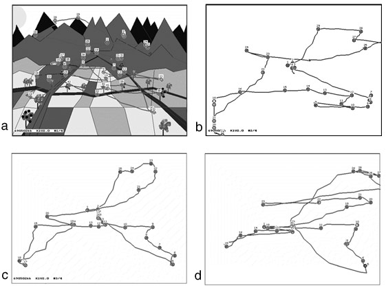
Illustration of a normal subject viewing a mountain landscape and the subject´s imagery, i.e. viewing the imagined picture on a blank screen: Saccade-fixation sequences, scanpaths (SP) are linked and numbered. First, in the real viewing phase (left upper SP) the landscape-picture is scanned for blue trees with a searchpath, after 5 seconds the first imagery of the picture is done (right upper SP), subsequently after 30 (left lower SP) and 60 (right lower SP) seconds imagery 2 and imagery 3. The similarity between viewing and imagery-SP and among the three imagery-SP is apparent: Obviously, a predictive top-down "plan" where to move the eyes governs the eye movement sequences of real and imagined scanpaths
(Joystone Gbadamosi and Wolfgang H. Zangemeister: Visual Imagery in Hemianopic Patients.J.Cognitivive Neuroscience 13 (7), 2001).
 |
|

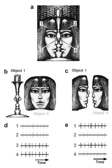
These figures illustrate that our perceptions depend to a crucial extent on the way our visual system segregates and binds features of complex scenes. The recognition of visual objects is preceded by a perceptual grouping operation that permits flexible and context dependent association of image features. One important goal of brain research is the identification of the neuronal mechanisms that accomplish these binding functions. This question has already been dealt with by Gestalt psychology, but there is still no consensus among neurobiologists concerning the neuronal mechanisms underlying these binding functions.
(Wolf Singer, Max Planck Institute for Brain Reserarch, 2001)
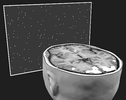 |
A subject views a scene consisting of stationary and moving light stimuli while being examined with functional magnetic resonance tomography. Continuously registered scans assess neuronal activity by measuring activity dependent upon changes in the oxygen content of blood. The visual cortex responds vigorously to moving stimulus arrays. (Lars Muckli, Rainer Goebel, Wolf Singer, Max Planck Institute for Brain Research "Cerebral activation following visual stimulation", 1999) |